Advanced Veterinary Diagnostics
East Lawn Animal Hospital is committed to providing your pet with the highest quality medical care possible. The advanced diagnostic imagery we use is essential to make sure we know everything we can about what is going on inside your pet. These tools offer non-invasive diagnostic methods that allow us to detect illnesses or abnormalities and provide better outcomes and a more precise prognosis for your pet.
What is an ultrasound?
Ultrasonography works by emitting high-frequency sound waves that travel through your pet. The sound waves bounce off internal organs or tissues to make echoes, which are then picked up and displayed as an image on an attached video monitor.
Ultrasonography is extremely useful in the examination process because images are displayed in real-time, allowing us to view both the structure and movement of internal organs.
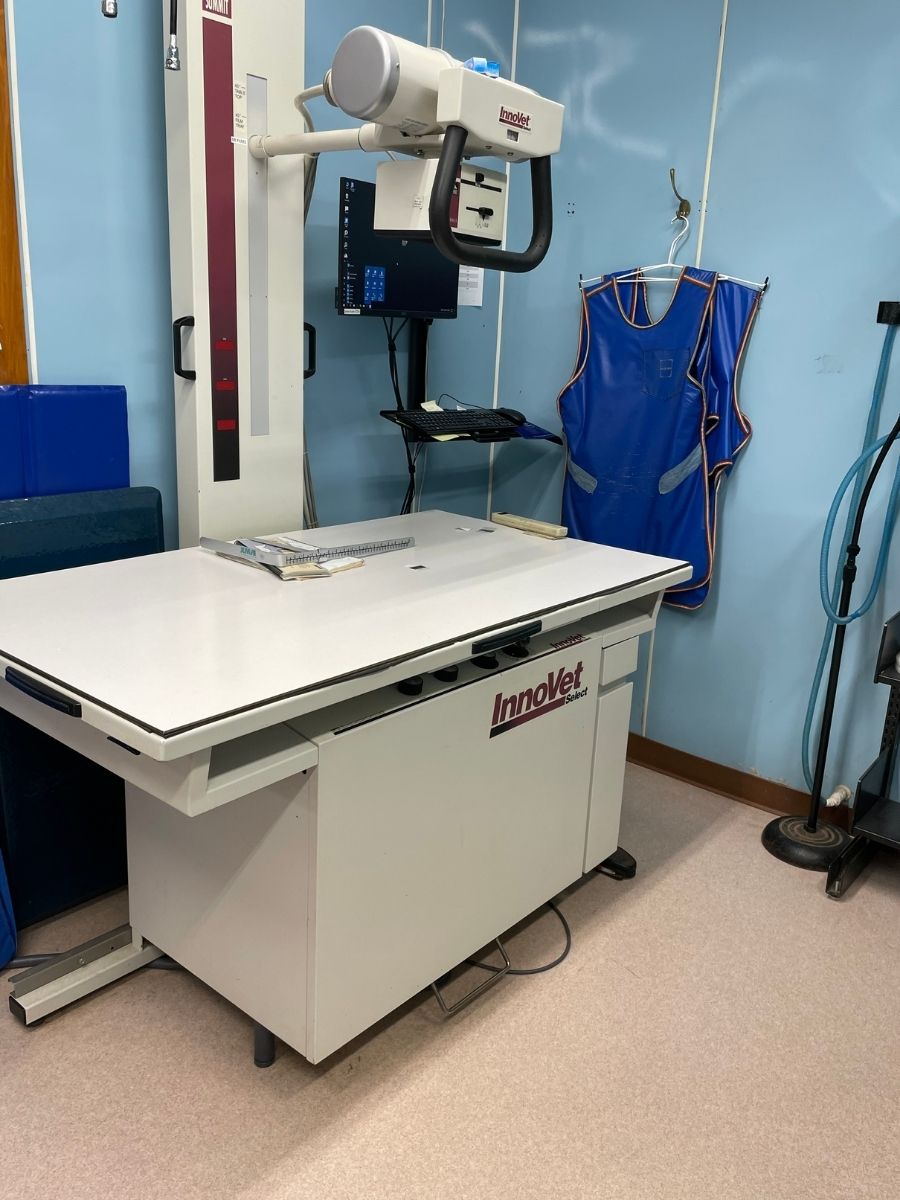
What is an X-ray?
A radiograph (X-ray) is a type of photograph that looks inside the body and reveals information that may not be discernible from the outside. Radiography can be used to evaluate your pet’s internal organs like the heart, lungs, and abdominal organs, as well as bones. When it comes to accurately diagnosing your pet, radiology can be an extremely valuable tool in our diagnostic arsenal. There have been many advancements in digital x-ray technology, and we can now manipulate the digital images that we take. This allows us to diagnose issues that may not be seen on a traditional x-ray.

